3-D Mammograms?
Are 3-D mammograms really better than the older kind? I’ve read that they find more breast cancer, but do they prevent deaths?
Andrew Weil, M.D. | March 24, 2023

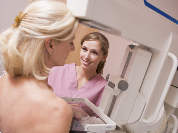
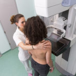
Digital breast tomosynthesis (what many refer to as 3-D mammography) was approved by the U.S. Food and Drug Administration in 2011, and it has since emerged as the new standard for breast cancer screening. Unlike original mammography, which provides a two-dimensional view of the breast, in tomosynthesis the scanner moves in an arc around the breast, capturing images from many different angles in order to create three-dimensional pictures. This provides a more complete view, especially in those with dense breast tissue. There is strong evidence that 3-D mammography can find more breast cancers, and at an earlier stage, than the older method can, and it has other benefits as well. Despite a few caveats, 3-D mammography is an excellent screening tool for detecting breast cancer – but we don’t know yet if it will reduce deaths from the disease.
Just a few years after the technology was approved for use in the United States, a 2014 study published in the Journal of the American Medical Association concluded that using the new 3-D technology in addition to digital two-dimensional mammograms led to the discovery of more breast cancers than digital mammography alone. Researchers reviewed the medical records of 281,187 women who had digital mammograms alone and compared them to the records of 173,663 women who had both types, and found that the combination turned up 5.4 breast cancers in every 1,000 women screened compared to 4.2 per 1,000 women found by digital mammography alone.
Since then, additional studies have confirmed multiple benefits of 3-D mammography:
- A reduction in “recall rate,” which is the number of women who need follow-up testing after a screening mammogram. Most of those who get called back after a mammogram do not have cancer, but it can be extremely stressful to be told the mammogram results require another look.
- Slightly fewer false results – both negative and positive – than older mammograms. False negatives are results that don’t find a cancer that’s present, leading to delays in diagnosis. False positives are results that identify a suspicious mass that turns out to be of no concern, but they create anxiety in women awaiting results and lead to unnecessary biopsies and surgeries.
- More significantly, fewer “interval cancers” (those cancers that emerge between routine screenings). A cancer that is found between screenings, usually due to the discovery of a lump or the development of other symptoms, tends to be a faster-growing, more dangerous tumor than one found on routine screening.
- Most important of all, 3-D mammograms detect more cancers overall, and at an earlier stage, than standard mammography.
It’s that last benefit that also comes with the caveat: Finding more cancers does not necessarily mean a reduction in mortality. A 2022 report from the nonprofit Lown Institute noted the risk of overdiagnosis and overtreatment of tiny, slow-growing cancers that may never progress to harm the patient.
In addition, a 2022 study found that contrast-enhanced mammography (CEM) was just about as accurate as digital breast tomosynthesis in identifying cancers. Although CEM received FDA approval in the same year as 3-D mammography, it has not been as widely adopted. CEM is sometimes used as an alternative to a breast MRI after a cancer has been identified, but a 2021 report in the journal Radiology noted that the procedure has the added risk of a poor reaction to the contrast injection, and it increases the patient’s overall exposure to radiation.
Since 3-D mammography and CEM are both still relatively new, we’ll need longer-term studies to show whether or not early detection saves lives, or simply generates more health care cost and patient anxiety as individuals treat cancers that might be better left alone.
Andrew Weil, M.D.
Sources:
Sarah M. Friedewald et al, “Breast Cancer Screening Using Tomosynthesis in Combination With Digital Mammography,” Journal of the American Medical Association, doi:10.1001/jama.2014.6095.
Johnson K, Lång K, Ikeda DM, Åkesson A, Andersson I, Zackrisson S. Interval Breast Cancer Rates and Tumor Characteristics in the Prospective Population-based Malmö Breast Tomosynthesis Screening Trial. Radiology. 2021 Jun;299(3):559-567. doi: 10.1148/radiol.2021204106. Epub 2021 Apr 6. PMID: 33825509. pubmed.ncbi.nlm.nih/33825509/
Siminiak N, Pasiuk-Czepczyńska A, Godlewska A, Wojtyś P, Olejnik M, Michalak J, Nowaczyk P, Gajdzis P, Godlewski D, Ruchała M, Czepczyński R. Are contrast enhanced mammography and digital breast tomosynthesis equally effective in diagnosing patients recalled from breast cancer screening? Front Oncol. 2022 Nov 24;12:941312. doi: 10.3389/fonc.2022.941312. PMID: 36505843; PMCID: PMC9730826. pubmed.ncbi.nlm.nih/36505843/
Jochelson MS, Lobbes MBI. Contrast-enhanced Mammography: State of the Art. Radiology. 2021 Apr;299(1):36-48. doi: 10.1148/radiol.2021201948. Epub 2021 Mar 2. PMID: 33650905; PMCID: PMC7997616. pubmed.ncbi.nlm.nih/33650905/
Originally Posted September 2014. Updated March 2022.


